


Hydrocele is a condition of advanced swelling of the scrotum with excessive fluid accumulation between the membranes covering the testicle. Normally, there is 0.5 to 1.0 ml fluid is present within this space to provide lubrication of the testicle. This fluid amount is 200 to 300 ml or more in hydrocele.
DIAGNOSIS
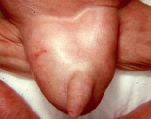
Because the appearance and patient history are very typical, diagnosis is quite easy. Scrotum seems swelled excessively unilaterally or bilaterally and tense. There is fluid in it.
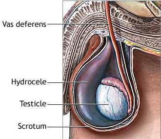


A typical elasticity sense is obtained when scrotum is hit by the finger during the examination. When scrotum is looked at by a light in a dark room, it seems pink. This simple examination called TRANSILLUMINATION finding proves that this swelling includes fluid and this is typical for HYDROCELE.
Simple hydrocele
Simple hydrocele is called stretched, oval shaped hydroceles which is unilateral, does not grow or shrink, is painless, may reach to very big sizes, has not a certain cause.


Secondary Symptomatic Hydrocele
It is generally occurs as a result of inflammations or tumors of the testicle or dependent structures within the scrotum in adult ages. Chronic hydrocele may develop in 10 to 15% of testicular tumors. Therefore, it should be controlled if there is any other disorder that accompanies with a scrotal ultrasonography in suspicious cases.
The only treatment for hydrocele is surgery.
HYDROCELECTOMY
Because to discharge the fluid by inserting a needle (PERCUTANEOUS ASPIRATION) tends to repeat and will cause cohesions for further operations, it should not be referred.

The illustration above shows a hydrocele sac which was liberated from the scrotum and removed from the cutline during the surgery. In the surgery, this sac is opened and discharged and sac leaves are reversed and sutured and placed into the scrotum.
SPERMATOCELE / EPIDIDYMIS CYST
They are small and painless masses located on and behind the testicle. It is a cystic formation including dead sperms.
A cystic formation occurs as a result of sperm accumulation. The cause is unclear. It does not cause pain. The patient notices an induration or swelling on the posterior upper side of the testis which is separate from the testis. It is diagnosed with examination and ultrasound. Treatment is not necessary unless it reaches to large volumes. It is removed by an operation if grows much.
